
|
By Helen Altonn A "second eye" is double-checking work of radiologists looking for early signs of cancer in mammograms at The Queen's Medical Center.
haltonn@starbulletin.comQueen's Breast Health Center is the first in Hawaii to use computer-aided detection (CAD) technology to help doctors detect hard-to-see breast cancers.
Kapiolani Medical Center for Women & Children also plans to install the system in the next couple of months.
Studies have shown the R2 ImageChecker, a CAD system approved by the Food and Drug Administration, increases early breast cancer detection by 23 percent.
"It's not the answer, but it's a useful adjunct to reading mammograms," said Dr. John Soong, Queen's radiologist.
Out of 450 to 500 mammograms that he read in the past 2 1/2 weeks, he said the ImageChecker spotted tiny calcifications concealed in dense tissue that he missed in two cases.
"That's the way it is supposed to work," he said. "I'm really starting to like this. ... If it is true that it picks up 23 percent more cancers, it is incredible for the patient."
|
Kathy Sugai, manager of Queen's Imaging Services, said mammograms are difficult to interpret because breasts can be dense and fatty and vary greatly."There are so many masses and calcifications. Little specks could evolve into something else. ... If we can detect this early enough, treatment is so sophisticated, the success rate is much higher."
It doesn't eliminate the need for mammogram screening or squeezing of breasts to get the best quality of images, Kristin Oishi, radiologic technologist, pointed out.
But it's a backup for tired eyes reading mammograms all day so radiologists "feel confident they haven't missed anything."
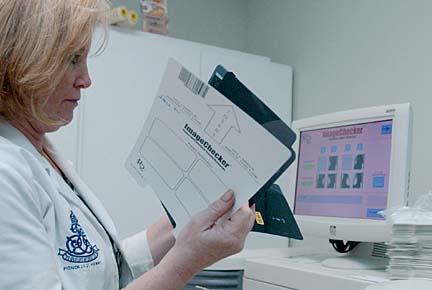
Showing how it works, Oishi loaded four mammogram films -- two views of each breast -- into the device similar to feeding paper into a fax machine. Within minutes it digitized and analyzed the mammograms, using a computer algorithm to spot and mark trouble spots.
It searches every pixel, looking for patterns that suggest microcalcifications or masses. "It's a very smart system," Oishi said. "It's fed all kinds of parameters, what to look for in cancer."
In the mammograms used for the demonstration, the ImageChecker flagged calcification areas in both breasts with triangles and marked a small mass with an asterisk on the left breast.
Soong had read the mammogram when it was taken a few weeks earlier and compared it with the woman's mammograms of the past couple of years. Comparing them to the computer images, he said, "I had checked those areas, but it is a helpful safety net."
Before the CAD was installed in late May, radiologists were doing double-reading mammograms to detect very early, subtle changes in the breast, he said.
"It's like playing a 'Where's Waldo?' game. If you don't know he's there, he's difficult to find. There may be one, four or no Waldos."
Soong said he is learning the ImageChecker's limitations and shifting his priorities to search for effects it cannot detect.
It is very good 98 percent of the time at marking calcifications and does not produce too many false negatives, he said.
But it isn't designed to look at some things, such as smooth masses and asymmetric densities, he said. They are not usually malignant, but they can be, he said, so he has put those at the top of his search list.
The ImageChecker detects about 80 percent of cancers, but doctors do better -- with an 89 percent detection rate, Soon said. Together, they push the detection rate up to 95 percent, he added.
R2 Technology Inc. generated great interest among Hawaii's health care professionals with a demonstration of its CAD system here in April.
Michelle Rudoy, manager of the Women's Health System at Kapiolani Medical Center for Women & Children, said, "We definitely think it's an important tool for increasing your detection rates ... and something we definitely want to offer our patients."
She said Kapiolani is working with Dr. Scott Grosskreutz of Hilo and submitting some proposals for reimbursements to the Hawaii Medical Service Association.
Grosskreutz, of Hawaii Radiologic Associates Ltd., East Women's Imaging Center and founder of the Hawaii Breast Cancer Society, is one of CAD's strongest Hawaii advocates.
He and other radiologists have been urging HMSA and other insurers to provide reimbursements to help health facilities acquire the advanced system, costing roughly $185,000.
Sugai said the Image- Checker is being used for all mammograms at no extra cost to patients, and Queen's has submitted its fees to insurers. "We've taken the position that since Medicare reimburses for it (for Medicare patients), we're fairly confident other insurance companies will follow," Sugai said.
HMSA Vice President Cliff Cisco said "benefits will be applied to CAD mammography," but no decisions have been made about the level of reimbursements.
He said the association's physician committees are reviewing the status of CAD mammography, how it relates to other procedures and if it will increase costs of care or reduce costs through early cancer detection.